
Page 4 - 2020 Spring CMTA Report
P. 4
The Surgical Correction of the CMT Foot:
IS SURGERY RIGHT FOR YOU?
PART 2 OF 4:
TENDON
TRANSFERS
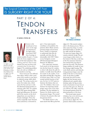
BY GLENN B. PFEFFER, MD Figure 1:
THE TIBIALIS ANTERIOR
elcome to the part 1. Four main muscles (figure #1). This muscle weakens
second part of our contribute to the problem: the early in the disease process. This is
series on the surgi- posterior tibial, tibialis anterior, what causes foot drop, or at least
cal correction of peroneus longus and peroneus diminished ability to dorsiflex at
the high-arched brevis. I think it is important the ankle and lift the forefoot.
(cavovarus) CMT to understand what they do The peroneus longus (figure #2)
W foot, focusing on normally, and what they do remains relatively strong. The per-
tendon transfers. In part 1, I wrote abnormally, with CMT. Videos oneus longus tendon inserts on
about the boney surgical correc- on my Instagram account the bottom of the foot, at the base
tion of the heel (calcaneus). Take (@Charcotmarietoothsurgery) of the first metatarsal (the bone
a look at your heel. Does it twist illustrate the difference. that extends from your big toe
Dr. Pfeffer is an ortho- inward? You were not born with First, some basic anatomy: joint into the arch). The peroneus
pedic foot and ankle
a turned-in heel. So how did it Each muscle in the leg connects longus stabilizes the inside of your
surgeon and co-director
happen? The underlying problem into the foot bones by a tendon, foot during weight bearing. The
of the CMTA’s Center
of Excellence at Cedars- has to do with an imbalance of so when I talk about the pull of a weak tibialis anterior and relatively
Sinai Medical Center, the muscles in your leg. tendon, I actually mean the pull of strong peroneus longus cause the
Los Angeles. He is also Some nerves are more affected the muscle on the tendon. A ten- inside of the foot to twist down-
a member of the
than others, which is what causes don looks and feels like a big piece ward. As the bone is pulled
CMTA’s Advisory
some of the muscles in your leg to of al dente pasta. With every step, downward, the arch becomes
Board.
be relatively strong, while others normal muscles function to bal- higher, the foot becomes more
are weak. The strong muscles over- ance the foot. In CMT patients, imbalanced and a painful callus
power the weaker ones, which however, this balance does not develops under the big toe joint.
causes the foot and toes to take exist. The imbalance that occurs Look at the bottom of your foot at
the abnormal twisted position so between two important muscle the base of the great toe. Many of
common with CMT. As a patient groups causes most of the major you will have this callus, caused by
with CMT grows during child- problems. The first imbalance is the increased pressure from the
hood and adolescence, the uneven between the tibialis anterior and abnormal position of the bone.
pull of the muscles on the devel- the peroneus longus. The second The second major muscle
oping bones can cause them to is between the posterior tibial and imbalance has to do with the pos-
become misshapen. Hence the the peroneus brevis. terior tibial tendon and the
need to surgically correct the heel The tibialis anterior is the peroneus brevis (figure #3). The
bone (calcaneus), as discussed in main muscle that lifts up the foot posterior tibial tendon connects to
4 THE CMTA REPORT SPRING 2020